What is 3D Printing in Anatomy?
3D printing in anatomy, also known as anatomical 3D printing, is a revolutionary technology that creates physical, three - dimensional models of anatomical structures based on digital data. It transforms digital medical images, such as those from CT scans, MRI scans, or ultrasound, into tangible objects.
The process begins with the acquisition of high - resolution medical imaging data. These images are then processed using specialized software. The software segments the relevant anatomical structures, separating them from the surrounding tissues. For example, if creating a 3D - printed heart model, the software will isolate the heart from the lungs, blood vessels, and other thoracic structures.
Once segmented, the digital model is sliced into thin cross - sectional layers. These layers are like the individual pages of a book, each representing a very thin slice of the anatomical structure. The 3D printer then reads these sliced layers and builds the physical model layer by layer. It deposits materials, which can range from plastics, resins, to even specialized biocompatible materials, in precise patterns according to the digital instructions.
For instance, in a 3D - printed kidney model, the printer will create the outer renal cortex, the inner medulla, and the complex network of blood vessels and ureters. Each layer is fused or bonded to the previous one as the printer builds upwards, gradually forming a detailed and accurate representation of the kidney's anatomy. This allows medical professionals, students, and researchers to hold and examine the model, providing a much more intuitive understanding of the complex internal structures than what is possible with just 2D images.
How Does 3D Printing Work in Anatomy?
Data Collection
The first crucial step in 3D printing for anatomy is data collection. The most common methods for obtaining anatomical data are through CT (Computed Tomography) scans and MRI (Magnetic Resonance Imaging). CT scans use X - rays to create cross - sectional images of the body. They can provide detailed information about bones, soft tissues, and internal organs. For example, a CT scan can detect small fractures in bones or identify tumors in the lungs with high precision. MRI, on the other hand, uses strong magnetic fields and radio waves to generate images. It is especially useful for visualizing soft tissues such as the brain, spinal cord, and muscles.
Accurate data collection is essential because any error or inaccuracy in the initial data will be carried through to the final 3D - printed model. A study by the Radiological Society of North America found that high - resolution CT scans with a slice thickness of less than 1mm can significantly improve the accuracy of 3D - printed anatomical models, reducing errors in the representation of fine anatomical details.
Model Creation
Once the data is collected, it needs to be transformed into a 3D model. This is achieved using specialized medical imaging software, such as 3D Slicer or Mimics. These software programs can import the raw data from CT scans or MRI and perform segmentation. Segmentation is the process of separating the different anatomical structures within the data. For instance, if creating a 3D model of the knee joint, the software will distinguish between the femur, tibia, patella, and the surrounding ligaments and tendons.
After segmentation, the model may need optimization and adjustment. This could involve smoothing the surfaces to make the model more anatomically accurate, filling in small gaps or holes in the data, and adjusting the overall geometry. The optimization process ensures that the 3D model is a faithful representation of the actual anatomical structure, ready for the printing process.
Printing Process
There are several 3D printing technologies available, each with its own advantages and applications in anatomy:
- FDM (Fused Deposition Modeling): This is one of the most common and affordable 3D printing technologies. FDM printers use a heated nozzle to melt and extrude a thermoplastic filament, such as PLA (Polylactic Acid) or ABS (Acrylonitrile Butadiene Styrene). In anatomy, FDM can be used to create large - scale models of bones or simple anatomical structures. For example, it can print a model of a human skull. The advantage of FDM is its low cost and ease of use, but it has relatively low precision, with layer thicknesses typically ranging from 0.1 - 0.4mm, which may not be suitable for very detailed anatomical features.
- SLA (Stereolithography): SLA printers use a laser to cure layers of photosensitive resin. This technology offers high precision, with layer thicknesses as low as 0.05mm. SLA is ideal for creating detailed models of small anatomical structures, such as the inner ear or a small blood vessel network. However, the resin materials used in SLA are more expensive, and the post - processing steps, such as cleaning and curing the printed parts, can be more complex.
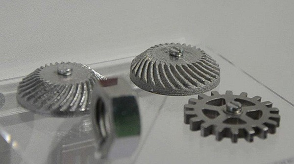
- SLS (Selective Laser Sintering): SLS printers use a laser to sinter powdered materials, such as nylon or metal powders. In anatomy, SLS can be used to create models with high strength and durability, like a functional model of a joint that can withstand mechanical stress. SLS has a high material utilization rate as the unsintered powder can be reused. But it has a relatively long printing time and the equipment is more expensive compared to FDM printers.
| 3D Printing Technology | Material | Precision (Layer Thickness) | Cost | Application in Anatomy |
| FDM | PLA, ABS | 0.1 - 0.4mm | Low | Large - scale bone models |
| SLA | Photosensitive resin | 0.05mm | High | Detailed small structures |
| SLS | Nylon, metal powders | Varies | High | High - strength functional models |
Yigu Technology's View
As a non - standard plastic metal products custom supplier, Yigu Technology holds a positive view on the application of 3D printing in anatomy. We believe that 3D printing in anatomy has a promising future. The ability to create customized anatomical models is a game - changer for medical education, surgical planning, and research.
However, we also recognize that there is still room for improvement in materials and technology. For materials, developing more biocompatible, durable, and cost - effective options is crucial. In terms of technology, enhancing the printing speed without sacrificing precision remains a challenge. Yigu Technology is committed to contributing to the development of 3D printing in anatomy by leveraging our expertise in material processing and manufacturing technology, aiming to provide better solutions for the medical industry.
FAQs
What are the common 3D printing materials used in anatomy?
Common 3D printing materials in anatomy include photosensitive resin and biocompatible materials. Photosensitive resin, like the ones used in SLA 3D printers, can achieve high - precision printing with smooth surfaces. It is great for creating detailed models of small anatomical structures such as the inner ear due to its ability to capture fine details with layer thicknesses as low as 0.05mm. Biocompatible materials, such as some types of polymers like PLA (Polylactic Acid) and PCL (Polycaprolactone), are suitable for models that may come in contact with biological samples or for simulating living tissues. They are non - toxic and do not cause adverse reactions in a biological environment, making them ideal for creating models for surgical training or tissue engineering research.
Is 3D printing accurate enough for surgical planning?
Yes, 3D printing is accurate enough for surgical planning. Many studies have demonstrated its high - level accuracy. For example, a 3D - printed model of the liver created from high - resolution CT scan data can closely match the actual anatomical structure of the liver. Research shows that the geometric accuracy of well - processed 3D - printed anatomical models can reach over 95% similarity to the real - life structures. In a case at a major hospital, a 3D - printed model of a patient's complex heart defect was used for surgical planning. The surgeons were able to accurately visualize the defect and plan the surgical approach on the 3D model. The success of the subsequent surgery, with the actual operation going smoothly according to the pre - planned strategy on the 3D model, further proves the reliability and accuracy of 3D - printed models for surgical planning.
How much does a 3D printer for anatomy cost?
The cost of a 3D printer for anatomy varies widely. Desktop - level FDM 3D printers, which can be used for simple anatomical model printing like basic bone structures, are relatively affordable, usually ranging from \(200 to \)2000. These printers are suitable for educational institutions on a budget or for initial research in small labs. Mid - range SLA 3D printers, known for their higher precision and better for detailed anatomical models, cost between \(2000 and \)10000. They offer better resolution and are often used in medical schools and research centers that require more accurate models. High - end industrial 3D printers, such as some SLS printers that can handle complex materials and large - scale, high - strength anatomical model production, can cost upwards of \(10000, even reaching \)50000 or more. Factors influencing the price include the type of the printer, its functions (such as multi - material printing capabilities), and the maximum print size.